
3D Cone Beam Imaging
Targeted 3D dental imaging to support diagnosis or treatment planning when clinically justified.
Information published by Dr Julia Musiol, dental surgeon in Antibes.
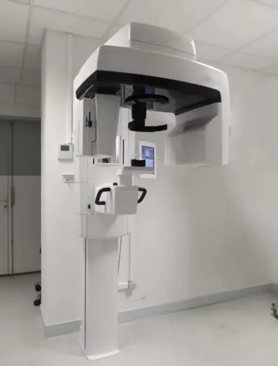
What is Cone Beam imaging?
Cone Beam computed tomography (CBCT) produces three-dimensional images of teeth and bone structures. Depending on the indication, it may be useful for implant planning, endodontics, impacted teeth or complex diagnostic situations.
The examination is not routine. The dentist first considers whether it will provide useful information that standard two-dimensional imaging cannot adequately supply. The field of view and exposure settings are tailored to the clinical question.
Procedure
You are positioned in the scanner while it rotates around your head. Images acquired from several angles are then reconstructed as a 3D volume. Tell the dental team if you are or may be pregnant before any X-ray examination.
Duration
The scan itself generally takes less than a minute. Positioning and checks bring the total appointment time to a few minutes.
Price
€69, subject to clinical indication and the estimate provided by the practice.
Preparing for your consultation
- Bring your prescriptions and mention relevant medication, allergies or medical history.
- If you have symptoms, note when they started and what seems to trigger them.
- When required, you receive a personalised treatment plan and written estimate.